
Ocular Manifestations of Hantavirus Infection: What Clinicians Should Know

Although the systemic manifestations of hantavirus infection are well recognized, its ocular effects are less commonly discussed. However, studies show that visual symptoms such as blurred vision and periorbital pain are relatively frequent, particularly in nephropathia epidemica (NE), the milder form of hemorrhagic fever with renal syndrome (HFRS) caused by Puumala hantavirus.
Recognition of these ophthalmic findings is important, especially during outbreaks, as ocular symptoms may occasionally precede systemic illness and prompt earlier diagnosis.
Myopia: The Most Common Ocular Finding
Transient myopia is the most frequently reported ocular manifestation in NE. Patients often present with sudden blurred distance vision caused by reversible refractive changes.
These changes are commonly associated with:
Lens thickening
Anterior chamber shallowing
Bilateral refractive shifts
Interestingly, visual acuity does not always worsen. Some hyperopic patients may temporarily experience improved vision during the acute phase of infection.
Fortunately, these refractive and anatomical changes are self-limited and typically return to baseline as the infection resolves.
Intraocular Pressure (IOP) Changes
The effect of hantavirus infection on intraocular pressure remains variable across studies.
Elevated IOP
Some reports describe acute angle-closure glaucoma during infection. Proposed mechanisms include:
Ciliary body edema or hemorrhage
Forward lens displacement
Zonular relaxation
Secondary inflammatory changes
Most cases resolve spontaneously, although some patients may require short-term anti-glaucoma treatment and cycloplegic therapy.
Reduced IOP
Other studies instead report decreased IOP during the oliguric phase of illness. This may result from transient reduction in aqueous humor production caused by ciliary body dysfunction, analogous to reduced renal filtration.
Because both hypotony and pressure spikes may occur, serial tonometry is recommended during the acute illness.
Eyelid Edema and Ocular Surface Findings

Several external ocular findings may occur even without specialized ophthalmic imaging. These include:
Eyelid swelling
Conjunctival chemosis
Hyperemia
Subconjunctival hemorrhage
Chemosis appears to be particularly common.
These findings are thought to result from hantavirus-induced endothelial dysfunction, leading to increased vascular permeability and plasma leakage. Importantly, these changes are usually transient and resolve during recovery.
Uveitis-Like Presentations
Whether hantavirus infection truly causes uveitis remains controversial.
Some case series have reported mild anterior uveitis that resolved spontaneously without treatment. However, larger prospective studies have failed to demonstrate definitive anterior or posterior uveitis.
Many investigators believe these findings may instead reflect transient vascular leakage involving the ciliary body rather than true intraocular inflammation.
Retinal Findings
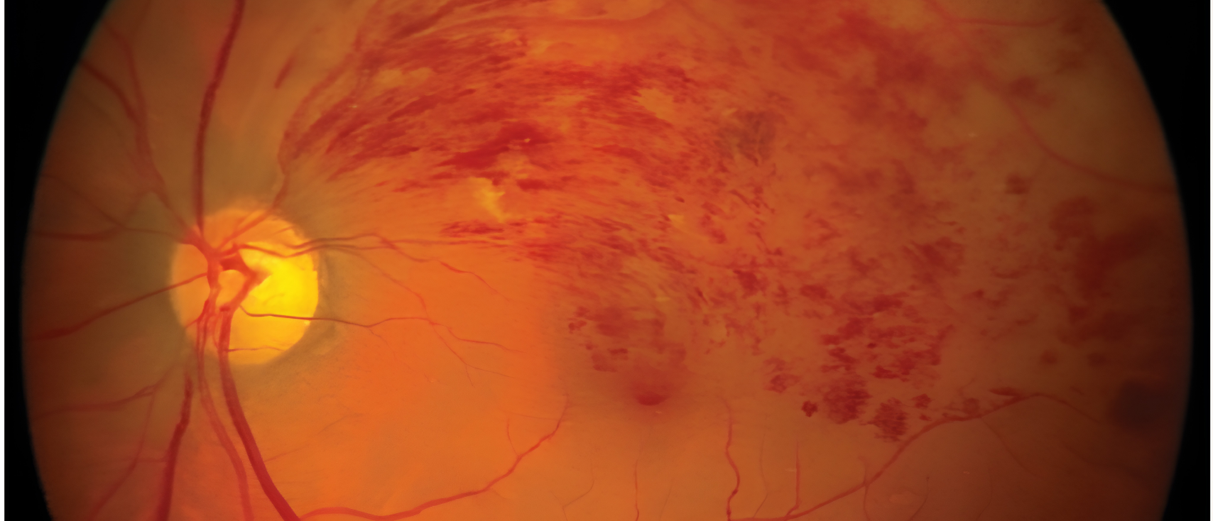
Retinal involvement in hantavirus infection appears uncommon but has been reported.
Documented findings include:
Retinal edema
Macular and peripapillary hemorrhages
Retinal vasculitis
Posterior necrotizing retinitis
Fundus examination in severe cases may demonstrate retinal whitening around the optic disc, flame-shaped hemorrhages, and venous sheathing.
General Evaluation and Clinical Implications
Although ocular complications are less common than systemic manifestations, recognizing these findings may facilitate earlier diagnosis and multidisciplinary management.
This is particularly important because ocular symptoms may occasionally precede systemic illness. Early identification of hantavirus infection may help reduce morbidity and mortality associated with systemic dissemination.
Ophthalmologists who suspect hantavirus infection should therefore consider appropriate systemic evaluation in addition to management of ocular complications. Timely treatment of the underlying disease may also help reduce ocular morbidity.
Key Takeaways
Transient myopia is the most common ocular manifestation of nephropathia epidemica.
Both elevated and decreased intraocular pressure have been reported.
Common anterior segment findings include chemosis, hyperemia, and subconjunctival hemorrhage.
True uveitis remains controversial.
Retinal involvement is rare but may include hemorrhages and vasculitis.
Recognition of ocular findings may aid earlier diagnosis of systemic hantavirus infection.
References:
Saari KM, Luoto S. Ophthalmological findings in nephropathia epidemica in Lapland. Acta Ophthalmol (Copenh). 1984;62(2):235–243.
Kontkanen M, Puustjärvi T, Kauppi P, Lähdevirta J. Ocular characteristics in nephropathia epidemica or Puumala virus infection. Acta Ophthalmol Scand. 1996;74(6):621–625.
Hautala N, Kauma H, Vapalahti O, et al. Prospective study on ocular findings in acute Puumala hantavirus infection in hospitalised patients. Br J Ophthalmol. 2011;95(4):559–562.
Saari KM. Acute glaucoma in hemorrhagic fever with renal syndrome (Nephropathia Epidemica). American Journal of Ophthalmology. 1976;81(4):455–461.
Baillieul A, Le TL, Rouland JF. Acute angle-closure glaucoma with choroidal effusion revealing a hantavirus infection. European Journal of Ophthalmology. 2021;31(1):NP4–NP8.